Forearm xrays are not able to fully assess the wrist joint or proximal radial ulnar joints and elbow joint.
Any suspected fracture with a dislocation/subluxation at the distal radial ulnar joint or proximal forearm dislocation/subluxation with the radiocapitellar joint or ulnohumeral joint
Consider ordering xrays of the wrist and elbow (the joints above and below) when you have high energy forearm injuries
In real life, xrays of the joints above and below an injury are not always ordered, but it is a good rule to remember especially in high energy injuries
Forearm xrays and other long bone xrays (humerus, femur, tibia/fibula) can be a great screening tool in polytraumas
Assess Quality: Forearm AP
Wrist is in AP position, with minimal overlap of the distal radius and ulna
The Radial styloid should be 180 degrees to the radial tubercle
Restoration of the radial bow in any radius open reduction internal fixation (ORIF) is the most important factor to restore
The radial bow allows for pronation, without it the radius could not overlap the ulna and this would severely limit a patient’s ability to pronate their forearm.
Perfect superposition of the capitellum and trochlea on the distal humerus (it looks like a perfect circle is made at the distal humerus when lined up perfectly)
Forearm xrays are not able to fully assess the wrist or elbow, but it is important to still evaluate these areas.
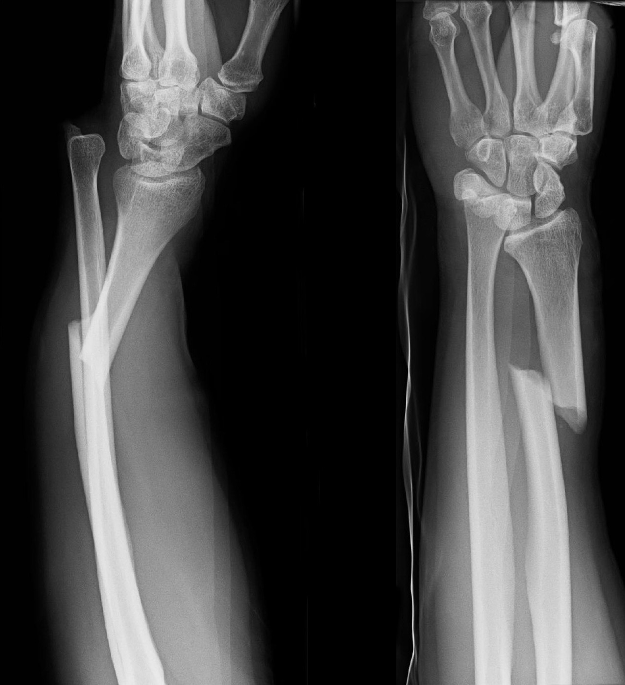
This is a cut off right forearm xray in a skeletally mature individual showing a distal third radial shaft fracture and distal radial ulnar joint dislocation.
This is also known as a Galeazzi fracture.
Order xrays of the ipsilateral wrist and elbow, at minimum the wrist is most important to assess with a dedicated wrist xray
Plan is closed reduction in the emergency room (ED), place in sugartong, board for open reduction internal fixation (ORIF) of the right radius with possible pinning of the distal radial ulnar joint
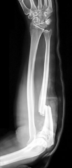
This is a trauma view of a right forearm (not perfect lateral), showing a proximal ulnar shaft fracture and radial head dislocation.
Also known as a Monteggia fracture.
Order xrays of the ipsilateral elbow and wrist, at minimum getting a dedicated elbow to visualized the radial head and rest of the joint more completely
Plan is for closed reduction in the emergency room with posterior arm splint with the arm in neutral. Board the patient for and open reduction internal fixation (ORIF) of the left ulnar with possible arthroplasty of the radial head (depending on if there are fractures present
Consider CT of the left elbow to better visualized the proximal radius and capitellum