How to Read Elbow Xrays
Elbow Anatomy
- This diagram demonstrates the area of the distal humerus that is the capitellum (articulates with the radial head on the lateral side) and trochlea (articulates with the proximal ulna on the medial side)
Lateral Elbow Xray
- The elbow series includes AP, External Oblique and Lateral
- A perfect lateral elbow xray must show the capitellum and trochlea aligned as shown by the red circle
- Other factors to assess are that the trochlear notch (thicker line), the radial head (thinner line) are articulating with the distal humerus.
- This is an example of a poorly done lateral, you can see that the distal humerus trochlea and capitellum do not line up because it doesn’t look like perfectly stacked circles
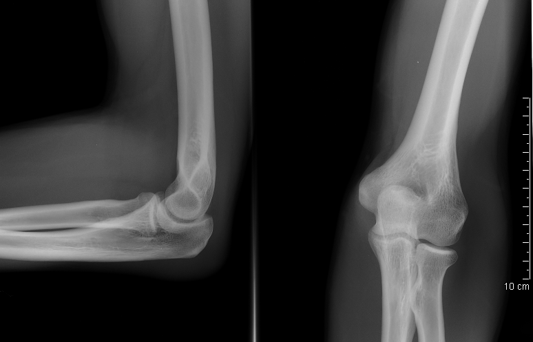
- The next parameter to assess is that the radius is always pointing to the center of the capitellum.
- The radius will always point to the capitellum on all views, except when there is subluxation or dislocation of the radiocapitellar joint.
- By MB - Collection personnelle, CC BY-SA 2.5, https://commons.wikimedia.org/w/index.php?curid=1254073
- As in all xrays you have to asses the whole image, even the soft tissue structures and each bony feature as well as to not miss anything
- Make sure to check the coronoid on the lateral as the radial head overlap can make it easy to miss
- This is what the coronoid looks like on a cadaver specimen, also review the medial and lateral liagments as well as the shape of the trochlea and capitellum.
- (Remember the radial head articulates with the capitellum and the ulna’s trochlear notch articulates with the trochlea)
- (https://shoulderelbow.org/2017/07/19/terrible-triad-elbow-fracture-dislocation/)
- The anterior and posterior fat pad signs can point to an occult fracture (one that cannot be seen on xray)
- The fat pad signs show up on xray often because there is bleeding into the elbow joint, this can come from a distal humerus fracture, a very proximal ulna fracture (olecranon fracture) or a radial head/neck fracture (the radial head/neck are intracapsular)
- The anterior fat pad is on the right side in the image below
- The posterior fat pad is on the left
- (https://www.aliem.com/emrad-ped-elbow/)
AP Elbow Xray
(https://radiopaedia.org/cases/normal-ap-elbow-radiograph)
- This is an AP of an elbow
- Ensure the radial head is pointing towards the center of the capitellum
- You can also use this view to check for the ulnohumeral (medial and left in image below) and radiocapitellar joint (lateral and right in image below) congruity as outlined in red.
- The joint should show equal spacing going from medial to lateral, if one side is widened this could mean that there is a ligament injury, entrapped fragment, or joint subluxation/dislocation.
- This same goes for the proximal radial ulnar joint as pointed to by the red arrow, spacing should be similar to the ulnohumeral and radiocapitellar joints if not slightly more narrow.
Oblique Elbow Xray
- The oblique view is mainly helpful to see another aspect of the cortices, which can be helpful to pick up hard to see xrays. Remember the same parameter applies of the radial head pointing towards the capitellum
Case example:
- Describe the fracture below
- How would you treat this?
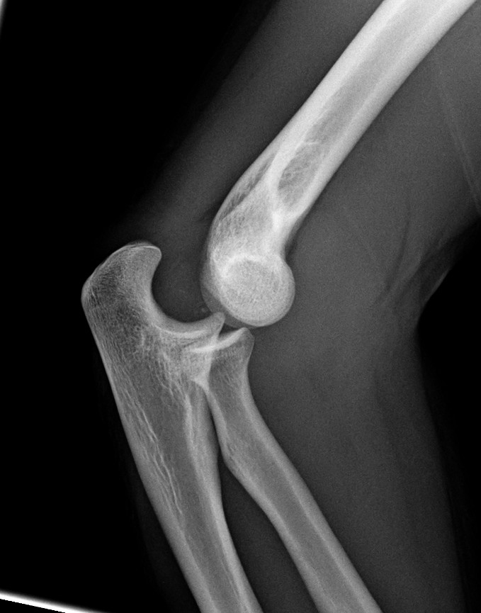
- This is an lateral and AP of a left elbow in an adult showing a simple posterior elbow dislocation
- Adult elbow because there are no physes visualized
- It is a simple elbow dislocation because there are no fractures
- Notice how the radial head no longer points to the capitellum in either the lateral or the AP
- It is a posterior dislocation because the proximal bone is always anatomically in the correct position
- Treatment steps
- Check patient for other injuries, consider ordering xrays of the ipsilateral humerus and forearm
- In real life, extra xrays are not always ordered, unless there is suspicion for further proximal or distal injuries. The clinical exam plays a factor and recognizing fracture types and how they occurred (high energy fractures for example are more likely to have more injuries and larger zones of injury).
- Perform a neurovascular exam
- Check skin to ensure the fracture is not open (if open always give an antibiotic and tetanus right away, commonly ancef in most cases)
- Perform a reduction (commonly done with sedation by the Emergency room team), extend the elbow and pull traction on the olecranon or distal forearm until you feel a reduction.
- Having the arm straight relaxes the triceps, allowing for the ulna and radius to be pulled distal allowing the distal humerus to sit in the joint.
- Ensure reduction with exam (smooth elbow range of motion, no deformity) and on fluoro (portable xray machine)
- Test your elbow stability by stressing the elbow with a valgus stress (testing the ulnar collateral ligaments, UCL) and a varus stress (lateral collateral ligaments, LCL).
- Then find where the elbow begins to subluxate when going from a flexed position to an extended position. If it dislocates or subluxates at 30 degrees, then note that in the record (saying, “stable from 30 to 130”) so the attending knows these parameters when the patient follows up in clinic. This helps the attendings to know the specific degree mark they become unstable so they can set a brace that will limit their motion so the patient does not get into a position that would allow for a dislocation to occur.
- Note that sometimes you have to keep the forearm in pronation or supination to keep the elbow fully reduced
- Pronation will use the intact UCL to lever up the lateral side of the joint
- Supination will use the intact LCL to lever up the medial side of the joint
- Place the arm into a posterior long arm splint (if very unstable, consider using stirrups to increase stability)
- Patient will be non weight bearing and follow up in 7-10 days.
- Check patient for other injuries, consider ordering xrays of the ipsilateral humerus and forearm