
How to Read Clavicle Xrays
When to Order Clavicle Xrays?
- When you suspect a clavicle fracture (Which seems obvious, but sometimes clavicle fractures can be easy to miss)
- Check under C-collars in high energy polytraumas, you just might find an open clavicle fracture!
- Whenever someone has pain over their clavicle, an xray is the first step
- Order bilateral AC joint xrays when there is AC joint involvement
- Can include shoulder series if severe to rule out shoulder injuries and type 4 AC joint injuries
- Order a serendipity view for suspected sternoclavicular dislocations
The Clavicle Series
- What should come in a typical 2 view clavicle xrays series?
- Upright AP xray
- This should be upright because gravity can show you the true displacement. Supine xrays may not show you how displaced the fracture is.
- Zanca view
- This is a 15degree upward or “Cephalic” tilt on the beam
- it shows you the clavicle without the scapula in the way and can give a better perspective on fracture displacement and morphology.
- This is a 15degree upward or “Cephalic” tilt on the beam
- Bilateral AC Joint view
- Not usually included in a standard clavicle series
- for AC separation injuries
- Serendipity View
- not in a standard clavicle series
- for SC joint dislocations
- Upright AP xray
Systematic Approach to Reading Clavicle Xrays
- Look at the whole AP and Zanca views and read what you see right away
- The clavicle is divided into three main areas:
- Medial Third: Includes the sternoclavicular joint
- Middle Third: Midshaft clavicle fractures
- Distal Third: Includes AC Joint as well as coracoclavicular ligaments
- Then zoom out and go through bony structures from proximal to distal.
- Sternoclavicular (SC) joint (it is easy to miss dislocations), compare to the other side if suspected. (Pain and/or deformity at the SC joint is very helpful to know)
- Outline clavicle cortices
- AC joint, Acromion
- Scapula
- Review all soft tissue from proximal to distal
AP Clavicle View
- Assess for this being the correct patient and side first
- The AP view shows the clavicle centered on the image and shows the whole bone from sternum to lateral acromion
- A weight bearing AP is of more use to truly assess fracture displacement
- Put in your xray order comments to have the patient upright when taking xrays
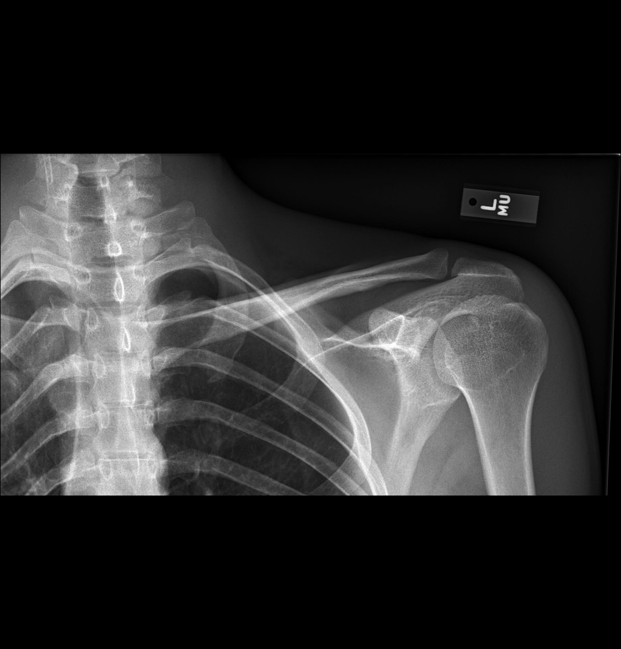

Cephalad View (Zanca View)
- This xray is taken with the patient upright as well
- Title of the beam is 15-20 degrees upward getting the scapula out of the way and showing a more true sense of the degree of fracture displacement and it gets the scapula out of the way
- In the image below the AP is at the top and the Cephalad view is the one below.
- Some surgeons call the cephalad view a “Zanca view”, others will say the Zanca has the xray image centered on the AC joint, not the full clavicle.
Zanca View (With AC jointed centered)
- This is a Zanca view, the image is aimed 15 degrees cephalad, and the AC joint is at the center of the image. (Some sources say that this is a true Zanca View)
- This image below is not adequate to assess the full clavicle
- For ortho purposes, a Zanca view or “Cephalad” view are used interchangeably and include the full clavicle at a 15 degree cephalad tilt.
Bilateral AC Joint View
- Bilateral AC joint views shows both AC joints, a stress view is considered to be with the patient standing upright as depicted in the lower image.
- As in other clavicle images, being upright can show you the true injury when gravity is acting on the upper extremity causing fracture or AC injury displacement, compared to when the patient is lying down
- (Researchgate.net)
Serendipity View
- The serendipity view is a bilateral clavicle view shot in 40 degrees of cephalad tilt (the Zanca view is 20 degrees and doesn’t include both clavicles)
- (Pacs.de)
- It is used to assess SC joint dislocations
- For posterior SC dislocations: The medial clavicle appears more inferior to the SC joint
- For anterior SC dislocations: The medial clavicle appears superior to the SC joint.
- (Orthobullets)
- The image below shows a serendipity view with a right posterior SC dislocation, it is subtle but the right SC joint is slightly incongruent to where it would be and also compared to the left SC joint.
- This is where your physical exam can be quite helpful in determining where the patient is painful
- Also, this is why if you have a suspicion, a CT is the next step in making the diagnosis
- (Emra.org)
Here is a CT of the same patient showing the right posterior clavicle dislocation setting just in front of the right subclavian vein and artery (yikes):
(Emra.org)
Sternoclavicular Dislocations (SC)
- They are usually anterior versus posterior
- Posterior SC dislocations are more concerning because the clavicle can push on a patients airway or cause damage to the great vessels underlying the sternum and SC joint.
- Obtain this imaging
- Serendipity view, 40 deg cephalic tilt that includes full bilateral clavicles
- AP Clavicle, upright
- CT Chest, to better eval location of dislocation
- There is no commonly used classification for SC dislocations.
Clavicle Classifications to Know
- There are no ubiquitously used clavicular shaft fracture classifications.
- When naming clavicle fractures it is best to start with saying whether they are “100% displaced”, “minimally displaced” or “nondisplaced” in the context of the location, medial 1/3, middle 1/3 or distal third 1/3. And whether there is an SC dislocation or AC separation.
- Why don’t they call AC separations, AC dislocations? Answer: I don’t know
AC Joint Classification
- To make an official diagnosis and grade of an AC joint separation you should have these three xray views
- AP clavicle, upright
- Zanca view (there is a letter C and A in the name to help you remember)
- Axillary shoulder view (required for type 4 AC separation to see if the clavicle is displaced posteriorly) Refer to: How to Read Shoulder Xrays for more info
- Rockwood classification
- Grade 1, reducible, coracoclavicular (CC) distance normal
- Grade 2, reducible, <25% CC ligament distance, CC ligaments still intact
- Grade 3, reducible, 25-100% superior separation of CC ligament distance, AC and CC ligaments are injured
- Grade 3A: no horizontal instability
- Grade 3B: Horizontal instability
- Grade 4, non-reducible, posterior dislocation of clavicle into trapezius muscle
- Grade 5, non-reducible, superior dislocation >100% CC ligament distance compared to contralateral, can cause skin tenting
- Grade 6, non-reducible, inferior dislocation of clavicle beneath coracoid
- (Wisc.edu)
- The Rockwood classification can be helpful to determine which type of AC separation requires surgery or not, Grade 4 and above or non-reducible separations usually require surgery to reduce the joint and stabilize it.
- Can consider surgery for type 3 injuries in laborers, elite athletes or cosmetic concerns or failed non op for chronic type 3 injuries.
Clavicle Surgical Indications
- Absolute indications
- open fractures
- displaced fracture with skin tenting subclavian artery or vein injury
- floating shoulder (clavicle and scapular neck fracture)
- Grade 4 and above AC separation
- Posterior SC dislocations with failed closed reduction
- with acute dysphagia, shortness of breath, decreased peripheral pulses
- Closed reduction under general with consideration for thoracic back-up for acute <3 weeks old posterior and anterior SC dislocations
- Relative and controversial indications
- 100% displaced with > 2cm shortening
- polytrauma patient
- bilateral displaced clavicle fractures
- brachial plexus injury (questionable because 66% have spontaneous return per Orthobullets)
- closed head injury
- TBI patients can have aggressive bone proliferation response in fracture healing, so I think about this as preventing larger amounts of bone formation with ORIF and shooting for primary bone healing instead of secondary bone healing
- seizure disorders
- grade 3 AC separations in laborers, elite athletes or cosmetic concerns or failed non op for chronic type 3 injuries
- chronic anterior or posterior SC dislocations that remain persistently symptomatic
- (Orthobullets)
Surgery Versus Non-Operative Discussion
- Fixing clavicles with a relative indication is debated. Many attendings will approach the discussion with patients from a shared decision making standpoint.
- More likely to fix young people, elite athletes, and/or heavy laborers with 100% displacement and >2cm of shortening
- Risks of nonunion or symptomatic malunion is higher with non operative treatment
- Decreased risk of nonunion with open reduction internal fixation with plates and screws (ORIF) treatment
- One surgeon’s approach for patients with relative surgical indications is to try non-operative treatment of displaced clavicle fractures for 10-14 days and then if the patient is able to forward flex and/or abduct their arm at or above their shoulder level there is a good chance that they could continue to be treated non-operatively. If there is significant difficulty with motion and ongoing severe pain, then the shared decision making becomes really important to help patients understand the risks and benefits of surgery versus no surgery.
- ORIF is more ideal if you can do it within 6 weeks, as fracture fragments are easier to mobilize and reduce.